Home
/ Anatomy Of Ribs And Chest : Bronchus | anatomy | Britannica / As with all parts of the body, the anatomy and physiology of the chest wall are intimately intertwined.
Anatomy Of Ribs And Chest : Bronchus | anatomy | Britannica / As with all parts of the body, the anatomy and physiology of the chest wall are intimately intertwined.
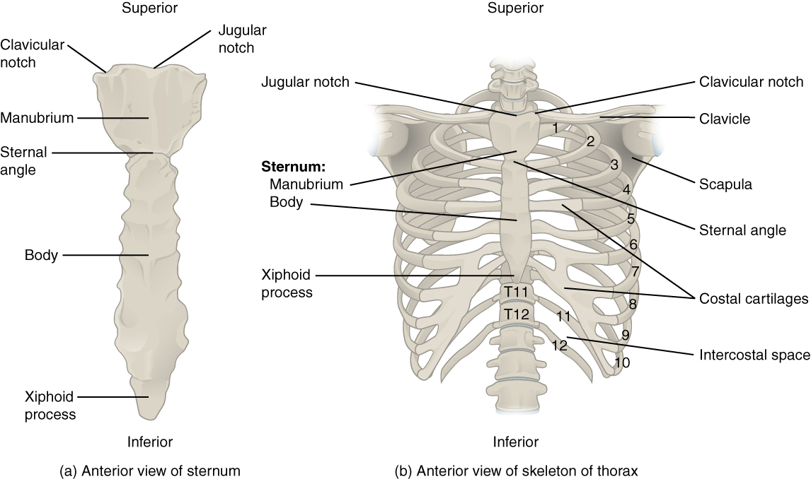
Anatomy Of Ribs And Chest : Bronchus | anatomy | Britannica / As with all parts of the body, the anatomy and physiology of the chest wall are intimately intertwined.. They also have a role in ventilation; External as i mentioned in my sternum anatomy video, the second pair of ribs meet at the junction. The first seven are connected behind with the vertebral column. Anatomy of the chest and the lungs: Basic rib anatomy consists of a head, neck, tubercle.
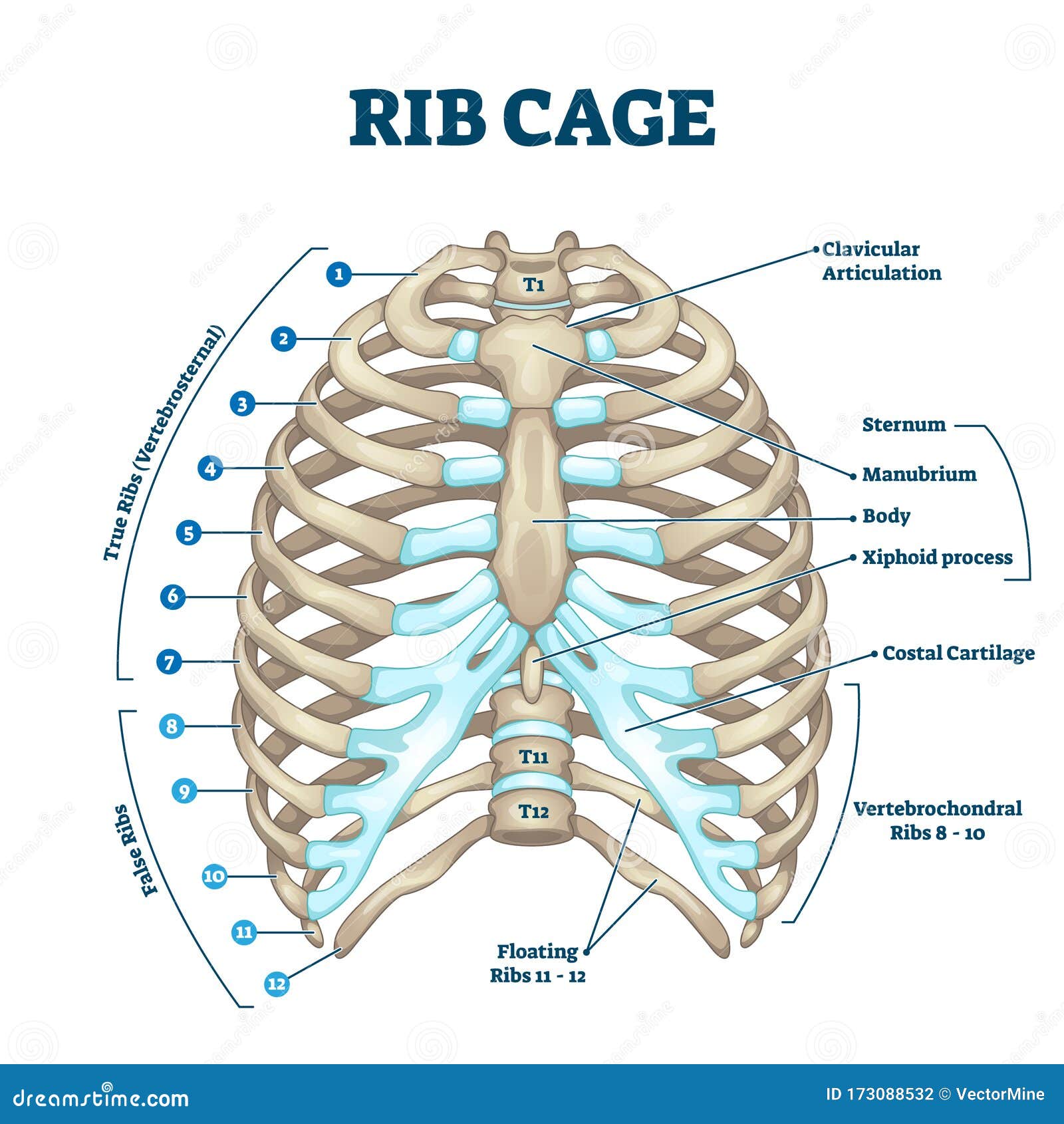
Right upper anatomy is to physiology as geography is to history: It extends from the position of the diaphragm to the clavicle or the collar bone. Ribs eight to ten are the false ribs and are connected to the sternum indirectly via the cartilage of the rib above them. The anatomical structure of the 24 ribs in the human body is complex because of the irregular shape and different lengths of each rib. They also have a role in ventilation;
7.4 The Thoracic Cage - Anatomy and Physiology from opentextbc.ca Ribs are divided into two basic groups: The first seven ribs attach to the sternum directly and are called true ribs. ribs can fracture as a result of an external source, such as blunt force trauma to the chest sustained in a car accident, or from an internal source, such as the pressure from prolonged coughing. Anatomy and physiology chest, ribs and respiratory system. True ribs, false ribs, and floating ribs. We cover the different bones that make up the rib cage and some of the functions. It describes the theatre of events. In this video we discuss the structure of the rib cage or thoracic cage. Ribs and other costal cartilage attach to it as will be examined in the following part of the article.
The sternum is a nearly flat rigid bone in the middle of human chest.
How these parts interrelate through joints is described also. O bones—spine, ribs, clavicles, scapulae, humeri. 12 photos of the anatomy of the chest. The embryologic and anatomic basis of modern surgery. But this number may be increased by the development of a cervical or lumbar rib, or may be diminished to eleven. Basic rib anatomy consists of a head, neck, tubercle. Anatomy of the chest and the lungs: The ribs stretches posteriorly from thoracic vertebrae the middle of every costal arch (being composed of a rib and its costal cartilage) with the exception in an anatomical position, the posterior end is higher and nearer the median plane in relation to the. To carry out the unique functions performed by. Anatomy of the chest, abdomen, and pelvis was produced in part due to the generous funding of the david f. The second most common chest wall abnormalities that we see on a cxr are metastases in vertebral bodies and ribs. The chest anatomy includes the pectoralis major, pectoralis minor and the serratus anterior. They also have a role in ventilation;
The sternum is a nearly flat rigid bone in the middle of human chest. Anatomy of the chest and the lungs: Right upper anatomy is to physiology as geography is to history: The ribs/costal cartilages have various attachments to the sternum. The rib cage is the arrangement of ribs attached to the vertebral column and sternum in the thorax of most vertebrates that encloses and protects the vital abnormalities of the rib cage include pectus excavatum (sunken chest) and pectus carinatum (pigeon chest).
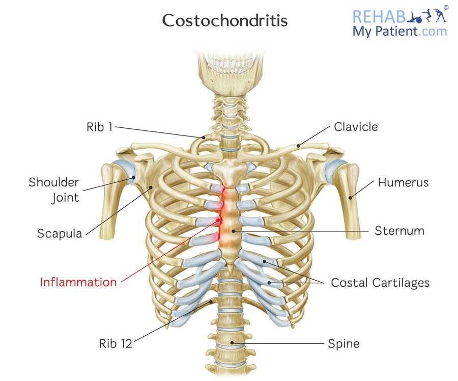
Costochondritis | Rehab My Patient from www.rehabmypatient.com Terms in this set (53). Ribs and other costal cartilage attach to it as will be examined in the following part of the article. They are twelve in number on either side; They are strong enough to support the skeleton and protect in this article, learn more about the number of ribs humans have, what their function is, and whether women have more than men. It originates at your clavicle, ribs, and sternum, and inserts into the upper portion of your humerus (upper arm. 620x465 px | 46 kb |703 views. Insert contains images of a typical rib and the first rib. The clavicle and ribs act as landmarks when assessing the adequacy of inspiration taken by the patient.
Anatomy and physiology chest, ribs and respiratory system.
Anatomy of the chest and the lungs: It discusses the specific anatomy of the ribs and costal cartilages, along with the sternum. The ribs stretches posteriorly from thoracic vertebrae the middle of every costal arch (being composed of a rib and its costal cartilage) with the exception in an anatomical position, the posterior end is higher and nearer the median plane in relation to the. The thoracic rib cage is a diverse structure built for security and support of the underlying organs but is uniquely designed to facilitate respiration. And as you might guess from the word major, it makes up the majority of the chest muscle mass. It extends from the position of the diaphragm to the clavicle or the collar bone. Anatomy and physiology chest, ribs and respiratory system. The anatomical structure of the 24 ribs in the human body is complex because of the irregular shape and different lengths of each rib. To determine if patient had good inspiration, what must be seen? Right upper anatomy is to physiology as geography is to history: The rib cage surrounds the lungs and the heart, serving as an important means of bony protection for these vital organs. Identify the following structures on the lateral chest radiograph: Ribs are divided into two basic groups:
Paschalides medical publications, 2004, with. True, false and floating ribs are denoted. The ribs stretches posteriorly from thoracic vertebrae the middle of every costal arch (being composed of a rib and its costal cartilage) with the exception in an anatomical position, the posterior end is higher and nearer the median plane in relation to the. It extends from the position of the diaphragm to the clavicle or the collar bone. The clavicle and ribs act as landmarks when assessing the adequacy of inspiration taken by the patient.
RIB - Rib (disambiguation) - JapaneseClass.jp from thumbs.dreamstime.com The sternum is a nearly flat rigid bone in the middle of human chest. As part of the bony thorax, the ribs protect the internal thoracic organs. How these parts interrelate through joints is described also. Insert contains images of a typical rib and the first rib. In this video we discuss the structure of the rib cage or thoracic cage. True, false and floating ribs are denoted. Terms in this set (53). Basic rib anatomy consists of a head, neck, tubercle.
List of nanda nursing diagnosis disturbed.
How these parts interrelate through joints is described also. The chest wall consists of 12 pairs of ribs, the first seven of which articulate both posteriorly with the spine and anteriorly with the sternum (figure 1 and figure 2). Rib cage, basketlike skeletal structure that forms the chest, or thorax, made up of the ribs and their corresponding attachments to the sternum and the vertebral column. Basic rib anatomy consists of a head, neck, tubercle. Spiral ct of thoracic inlet. Identify the following structures on the lateral chest radiograph: Chest blunt trauma (cbt) and the resultant rib fractures often lead to thoracic collapse. As part of the bony thorax, the ribs protect the internal thoracic organs. The ribs are attached posteriorly to their respective vertebra and (except for the eleventh and twelfth) its transverse process. Right upper anatomy is to physiology as geography is to history: Ribs eight to ten are the false ribs and are connected to the sternum indirectly via the cartilage of the rib above them. It discusses the specific anatomy of the ribs and costal cartilages, along with the sternum. In this video we discuss the structure of the rib cage or thoracic cage.
620x465 px | 46 kb |703 views anatomy of ribs. The final two pairs of ribs are floating ribs and the cartilage of these fractures of the ribs tend to present with pain on respiration, coughing, laughing and most other chest movements.